Don’t judge a book by its cover, they said, but pathology says, you can learn a human or animal, by examining the components of its body and nature –including the epidermis, dermis, and hypodermis.
Pathology is the scientific study and diagnosis of disease by examining the body’s organs, tissues, bodily fluids, and even the whole body. It involves analyzing blood, urine, stool, and other bodily tissues to identify any abnormalities that may indicate the presence of a disease, including cancer, chronic illnesses, and health risks.
Pathology is comprised of four main specialties, namely –
- Anatomic pathology,
- Dermatopathology,
- Forensic pathology, and
- Laboratory medicine.
Each of these specialties has its own expertise and limitations, including low accuracy of diagnosis, delayed delivery time, and high costs associated with obtaining, managing, and processing pathological data. As a solution to these challenges, Digital Pathology was developed, which not only offers improvements in diagnostic accuracy and turnaround time but also enables applications such as teaching, research, and primary diagnostic reporting.
What is Digital Pathology and How is Digital Technology and Collaborative Partnerships Revolutionizing Pathology?
Digital pathology is the process of using digital technology for simplifying and optimizing the acquisition of pathological data –via digital scanners, management, sharing, and interpretation of information related to pathological examinations and testing, saved in the form of cloud data, slides, articles, or short notes in a digital environment or system. It leverages computer-based technology such as virtual microscopy, which converts glass slides into digital slides that can be viewed, managed, analyzed, and shared on a computer monitor.
The 10 Best Companies in Digital Pathology Market 2023, have identified several unique reasons why digital pathology is becoming increasingly popular in the medical field:
- Improved accuracy and efficiency: Digital pathology eliminates the need for pathologists to physically examine tissue samples under a microscope, which can be time-consuming and prone to human error. Digital images can be quickly and accurately analyzed using computer algorithms, leading to faster and more accurate diagnoses.
- Remote consultation and collaboration: Digital pathology allows pathologists to share images and collaborate with colleagues in different locations, which can be especially helpful in rural or remote areas where access to specialists may be limited.
- Better record-keeping: Digital images can be easily stored and retrieved, making it easier to track patient histories and monitor disease progression over time.
- Improved education and training: Digital pathology provides a more accessible and standardized way to train new pathologists, as they can study and review digital images from a wide range of cases.
- Minimal waiting times: Digital pathology allows for faster turnaround times in diagnosis, as images can be processed and analyzed more quickly than traditional methods. This can reduce the time patients spend waiting for a diagnosis and receiving treatment.
- Improved precision of diagnosis: Digital pathology allows for more accurate and precise diagnoses, as pathologists can zoom in and manipulate digital images to examine tissue samples in greater detail. This can help to identify smaller or more subtle abnormalities that may be missed with traditional methods.
- Swift treatment: With faster and more accurate diagnoses, patients can receive treatment more quickly, potentially improving their outcomes and quality of life.
- Better pathologist efficiency: Digital pathology can improve the efficiency of pathologists, as they can review and analyze images from anywhere with an internet connection. This can reduce the time and resources needed for travel and on-site consultations.
- Cost-effective returns on external consultations: Digital pathology allows for easier and more cost-effective external consultations with specialists, as images can be shared and reviewed remotely. This can help to reduce the need for expensive travel and on-site consultations.
- Minimized cost of handling glass slides internally: Digital pathology eliminates the need for storing and managing physical glass slides, which can be costly and time-consuming. Digital images can be easily stored and retrieved, reducing the need for physical storage space and resources.
- Infrastructure savings such as in laboratory and office spaces: Digital pathology can reduce the need for physical laboratory and office spaces, as pathologists can review and analyze images remotely. This can help to reduce overhead costs and increase efficiency.
Overall, digital pathology has the potential to improve diagnostic accuracy, speed up diagnosis times, and enhance education in the medical field and encourage beneficial collaboration between human pathologists and robotic pathologists.
Exploring the Future of Pathology: Integrating Human Expertise with Robotic Pathology Technology
The future of pathology is somewhat complicated, as the number of cases that require the attention of pathologists is increasing, while the practicing pathologists are retiring at a faster rate than the current replacement rate. Despite the promising advancements in AI technology, the major challenge facing pathology remains difficult to overcome. This is due to a limited availability of qualified pathologists, and the reluctance of some human pathologists to collaborate with robotic counterparts, making it challenging to bridge the gap. But if the collaboration is achieved amicably, it will redefine innovation in pathology.
For Example:
- Whole slide imaging (WSI): A digital imaging technique a microscope under robotic and computer control is used in pathology that allows the entire tissue sample on a glass slide to be scanned and converted into a high-resolution digital image. This technology enables the creation of a digital slide that can be viewed and analyzed on a computer monitor, rather than through a microscope. WSI uses specialized scanners to capture images of the entire slide, and the resulting digital image can be stored, analyzed, and shared electronically. This technique has many applications in pathology, including primary diagnosis, research, education, and telepathology.
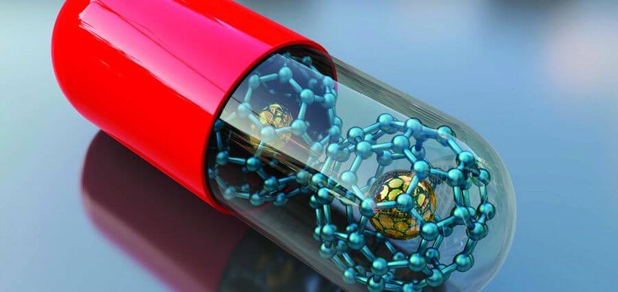
- Nanotechnology: Nanotechnology in pathology involves the manipulation and assembly of nanoparticles, nanomaterials, and other nanoscale devices to detect, diagnose, and treat diseases at the molecular and cellular level – a perfect application for industrial robotics. Nanoparticles can also be used to enhance imaging techniques, such as magnetic resonance imaging (MRI) or computed tomography (CT), allowing for more accurate and detailed imaging of tissues and organs. Nanotechnology can be used to develop biosensors that can detect and diagnose diseases in real-time. Biosensors can be designed to detect specific biomarkers or molecules in bodily fluids, such as blood, urine, or saliva, and provide rapid and accurate diagnoses. Nanoparticles can also be used to create advanced diagnostic tools, such as microfluidic chips, that can analyze and process samples more efficiently and accurately than traditional methods.
So, rather than being concerned about robots replacing human pathologists, we should encourage the partnership and cooperation between human pathologists and robotic counterparts, which can be seen as a ground-breaking development in the field of pathology.
-by Chidiebere Moses Ogbodo
Read More Articles: Click Here


